avm classification radiology
Its utility is based on accurate initial diagnosis that correlates consistently with clinical presentation disease course and treatment. We describe the various classification systems which have been devised to define the multiple.

Radiology Classification Of Vascular Malformations
3 This classification has four stages with stage 1 lesions being asymptomatic and stage 4 representing high-output heart failure.

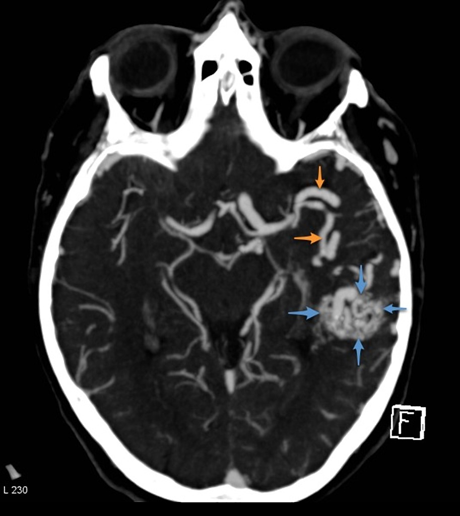
. A nidus is detectable on catheter angiography by being visible on both arterial and venous phases of circulation. An appropriate classification scheme for vascular anomalies and definite indications for treatment are important to successful treatment overall. And Interventional Radiology 28730All India Institute of Medical Sciences New Delhi India.
As information has been gleaned a new classification system has emerged that divides vascular anomalies into neoplasms and malformations. Magnetic resonance MR imaging is the most valuable modality for classification of vascular anomalies because it accurately demonstrates their extension and their anatomic relationship to adjacent structures. Vascular malformations like arteriovenous malformation AVM dural arteriovenous fistulas dAVF aneurysms cavernoma DVA very rare.
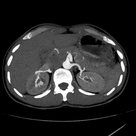
While the treatment of most vascular anomalies is multifactorial interventional radiology procedures including embolic therapy sclerotherapy and laser. The single feeder artery was from the distal abdominal aorta originating below the inferior mesenteric artery. The AVM was located within the lower pole of the left kidney.
Peripheral vascular malformations are now described according to some accepted guidelines and the principle of proper treatment nidus ablation is becoming clear. Frieden I Enjolras O Esterly N. Benign infantile hemangiomas represent the vast majority of vascular tumors and occur most commonly in their sporadic form.
A comprehensive assessment of vascular anomalies requires functional analysis of the involved vessels. 1 Pulmonary AVMs may be classified as simple with a single feeding and draining vessel 80 of cases or complex with 2 or more feeding or draining vessels 20 of cases. Classification diagnosis and interventional radiologic management of vascular malformations.
The Yakes AVM Classification System is utilized to determine endovascular approaches and the embolic agents that will be successful to ablate these AVMs. Crossref Medline Google Scholar. Although transcatheter embolization is a less-invasive and effective treatment option it has a potential risk of complications including renal.
The Schobinger AVM Classification for peripheral AVMs non-neuro is useful to quantify the degrees of symptomatology a patient possesses regardless of the AVMs angioarchitecture 10. 80 1 Or into four types 2. Renal arteriovenous AV shunt a rare pathologic condition is divided into two categories traumatic and nontraumatic and can cause massive hematuria retroperitoneal hemorrhage pain and high-output heart failure.
Up to 65 of pulmonary AVMs are found in the lower lobes of the lung. Orthop Clin North Am 2006373435474 viiviii. The Schobinger classification is a clinical assessment of vascular shunting that is predictive of treatment success Table 2.
Infarction with hemorrhagic transformation Hemorrhagic venous infarction in sinus thrombosis Hemorrhagic primary brain tumors or metastases Drug abuse PRESS Reversible cerebral vasoconstriction syndrome. Spinal arteriovenous malformations can be classified in a number of ways. Their frequency varies occurring in roughly 10 to 20 persons per 100000.
Vascular tumors are further classified as benign locally aggressive or malignant. Intramedullary glomus AVM type. The term vascular anomaly represents a broad spectrum of vascular pathology including proliferating vascular tumours and vascular malformations.
The most recent classification scheme of 2014 continues to divide vascular anomalies into vascular tumors and vascular malformations. Catheter angiography of renal AVM. 101055b-0039-172052 5 Cerebral Arteriovenous Malformations AVMs and Dural Arteriovenous Fistulas dAVFs 51 Evaluating AVM Angioarchitecture 511 Clinical Case A 47-year-old female with new s.
There are several subgroups the most common being glomerular type brain AVMs with fistulous type AVMs being less common. Hemangioma and arteriovenous malformation AVM. Brain arteriovenous malformations AVMs are abnormal vascular connections within the brain that are presumably congenital in nature.
Vascular birthmarks and other abnormalities of blood vessels and lymphatics. Single coiled vessel spinal dural AV fistula type II.

Endovascular Treatment Of Arteriovenous Malformations Of The Head And Neck Focus On The Yakes Classification And Outcomes Journal Of Vascular And Interventional Radiology

Pdf Classification Diagnosis And Interventional Radiologic Management Of Vascular Malformations Semantic Scholar
The Radiology Assistant Non Traumatic Intracranial Hemorrhage
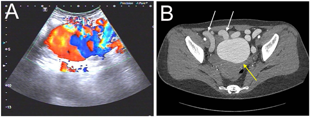
Frontiers Case Report A Rare Abdominopelvic Arteriovenous Malformation Originating From Splenic Artery And Draining Into Portal Vein

Pdf Peripheral Arteriovenous Malformations Imaging And Endovascular Management Strategies
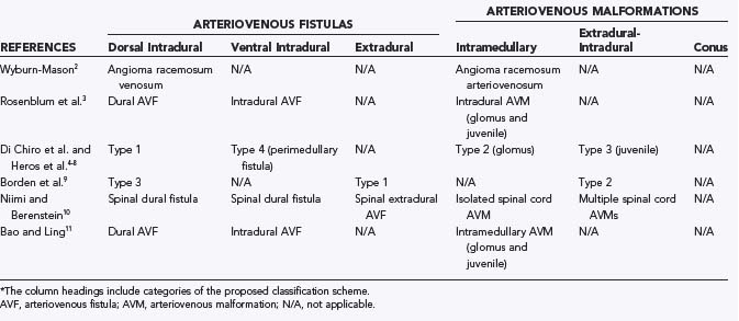
Classification Of Spinal Arteriovenous Lesions Clinical Gate
Arteriovenous Malformation Radiology Reference Article Radiopaedia Org
Cerebral Pial Arteriovenous Malformation Avm

Vascular Malformation Classification References T M O Couto Download Scientific Diagram

Vascular Anomalies Of The Head And Neck Diagnosis And Treatment Springerlink
Imaging In Neurovascular Conflict Of The Trigeminal Nerve With Grading Of Its Severity Semantic Scholar

Yakes Avm Classification System 8 Download Table

Vascular Malformation Classification References T M O Couto Download Scientific Diagram

Issva Classification Of Vascular Anomalies A Simple

Epos
Arteriovenous Malformation Cerebral Radiology Case Radiopaedia Org
Renal Arteriovenous Malformation Radiology Reference Article Radiopaedia Org

Hamburg Classification System Of Vascular Malformations Radiology Reference Article Radiopaedia Org

What Are Vascular Anomalies Ucsf Radiology
0 Response to "avm classification radiology"
Post a Comment